
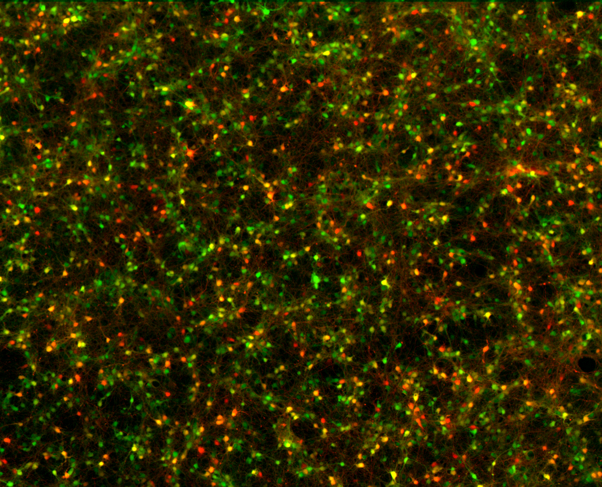
At about one in the morning on August 4, 2004, Edward Boyden put a dish of cultured neurons under a microscope at Stanford and directed pulses of blue light at a cell. The neuron fired. Its electrical spikes followed the flashes closely enough to suggest that light could become a precise experimental command. Boyden later described the night in a first-person scientific account: after hours of recording, he emailed his collaborator Karl Deisseroth, “Tired, but excited.”[2]
There had been practical reasons to doubt that the experiment would work. A light-sensitive protein borrowed from an alga had to function in the very different surroundings of a mammalian neuron. Boyden’s account records months of preparation, including work on the optical equipment and methods for putting the protein into cells. The overnight result gave the collaborators a way to pursue the idea with direct electrical measurements.[2]
On Monday, the Nobel Assembly awarded the 2026 physiology or medicine prize to Deisseroth, Peter Hegemann and Georg Nagel for discoveries concerning light-gated ion channels and optogenetics. The award recognizes a chain of discoveries that connected microbial biology to experiments on the living brain.[1]
The attraction is easy to understand. An electrical stimulus can excite neighboring cells as well as the intended ones; a drug may act across a wide area and linger. Researchers needed more control over which cells they affected and when. Optogenetics joined genetic selection to the speed of light pulses, giving them a more discriminating way to intervene in a circuit and watch what followed.[3]
A useful protein in an unlikely place
For Hegemann and Nagel, the route into neuroscience began with a difficult biochemical problem. The algal photoreceptors they wanted to study were scarce, unstable and hard to purify. In their later account, the breakthrough in approach came when Suneel Kateriya in Hegemann’s group identified promising DNA sequences in a Chlamydomonas database in 2001. Instead of extracting enough of the fragile material from algae, the researchers could ask another cell to make it.[4]
Nagel brought experience in studying membrane proteins inside frog egg cells. Those large cells provided a setting in which the team could introduce genetic instructions and measure the resulting electrical behavior. The collaboration made a small organism’s response to light accessible to a different kind of experiment: the researchers could investigate the protein separately from the alga that normally used it. The approach also depended on retinal, the small molecule that absorbs light within these proteins. Mammalian brain tissue already contains it, helping the transferred protein function without requiring a separate supply of that ingredient. This biochemical compatibility helped make the method practical.[4]
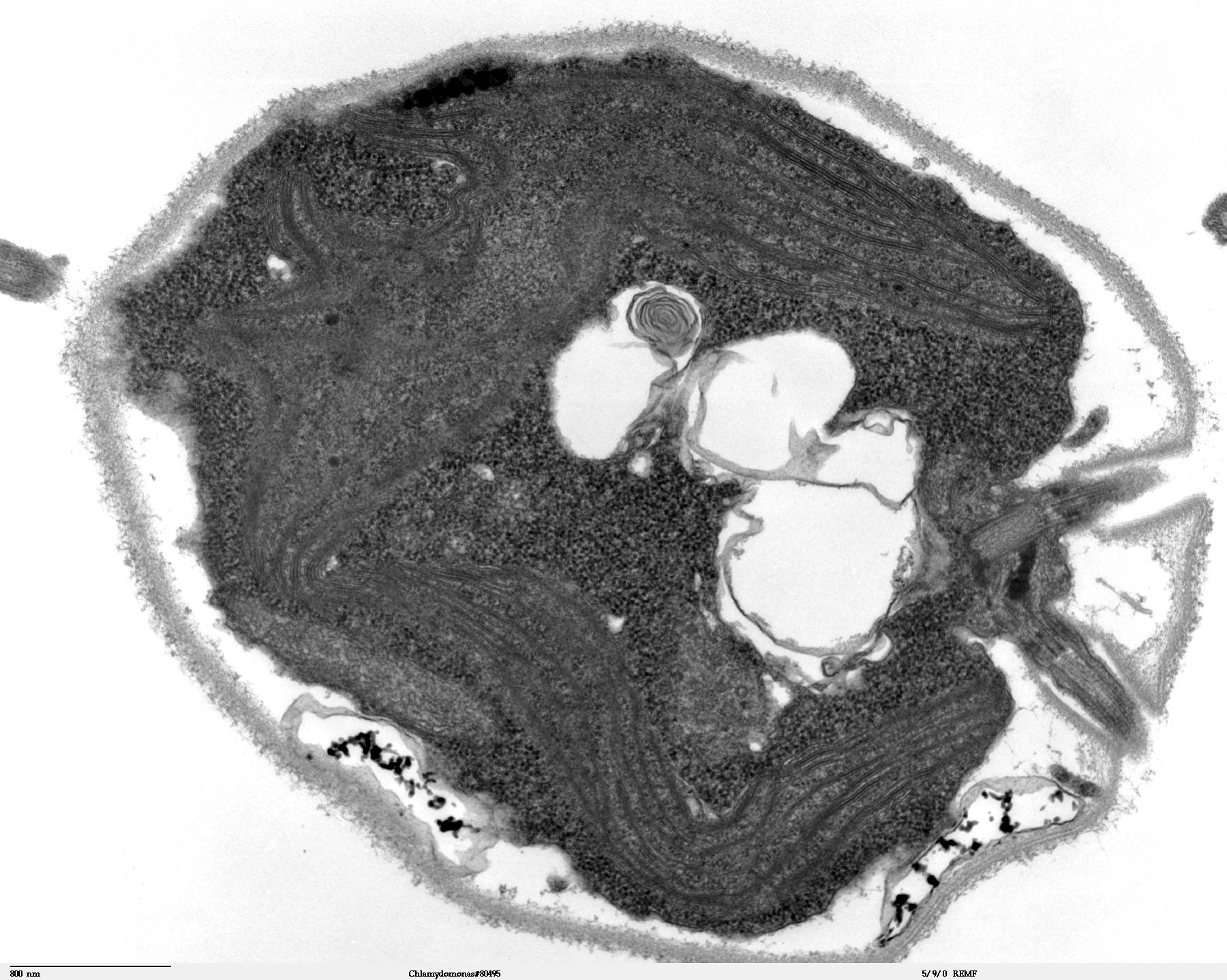
Image details
Original file; no crop, padding, color adjustment or depicted-content alteration.
Their 2002 Science paper identified channelrhodopsin-1. When the relevant genetic sequence was expressed in frog egg cells, with the light-absorbing molecule retinal present, illumination produced an electrical conductance selective for protons. Even the protein’s central membrane-spanning region could do the job. The finding placed the light response in a compact molecular structure that could operate outside its original organism. The authors proposed a role in the green alga’s movement in relation to light, known as phototaxis.[5]
A second protein made the opportunity more striking. In 2003, Nagel, Hegemann, Ernst Bamberg and colleagues reported that channelrhodopsin-2 worked as a directly light-gated channel in frog egg cells and mammalian cells. It admitted positively charged ions when illuminated. Opening such a path changes the voltage across a cell membrane: ions flow through the channel, carrying charge with them.[6]
The protein combined two jobs that might otherwise require a longer chain of molecular events. It detected light and provided the route across the membrane. Its response was also dynamic. During continuous illumination, the initial current weakened toward a smaller steady level; after a pause, sensitivity recovered. Those properties mattered because a useful experimental switch would have to be characterized over repeated pulses, not merely shown to respond once.[6]
The 2005 neuron paper, by Boyden, Feng Zhang, Bamberg, Nagel and Deisseroth, demonstrated what that switch could accomplish in mammalian nerve cells. The researchers used a viral delivery method to introduce the gene and rapid optical switching to stimulate the resulting protein. They could control firing on a millisecond timescale and evoke both excitatory and inhibitory synaptic transmission.[7]
That last distinction is useful: a neuron’s electrical spike is an event inside one cell, while synaptic transmission is how its activity influences another. A tool that could time those events allowed experiments to follow communication through a circuit. Light pulses became a way of specifying when selected cells should contribute, with a precision suited to the signals being studied. The published work established the technique through measurements of spikes and synaptic events, rather than a claim that illuminating a brain would reveal its workings automatically.[7]

Image details
Wikimedia proportionally reduced rendition of the original portrait; no additional crop, padding or depicted-content alteration. Image retains CC BY-SA 4.0.
Choosing what to change
An activating switch supplied only part of the experimental toolkit. In 2007, Zhang and colleagues developed a light-driven chloride pump from an archaeal microorganism, Natronomonas pharaonis, to suppress neural firing. It could prevent individual spikes or maintain inhibition for longer periods. Because the pump and channelrhodopsin-2 responded to different parts of the light spectrum, they could be combined for opposing effects.[8]
Being able to increase and decrease activity widened the questions an experiment could ask. Activation could test whether a selected population was sufficient to produce an effect; suppression offered a way to investigate what depended on that activity. The 2007 work demonstrated the complementary tools in neural tissue and used them to control movement in roundworms. It was a platform for carefully designed interventions, with the biological question still determining which cells to target and what to measure.[8]
One of the early demonstrations in a living mammal concerned waking up. Antoine Adamantidis, Zhang and colleagues targeted neurons that produce hypocretin, also called orexin, in mice. Those cells were already implicated in maintaining stable arousal. The unresolved question was whether their firing could help cause a transition out of sleep, rather than simply accompanying it. An optical fiber let the researchers stimulate the selected population deep in the brain while the animals could move freely.[15]
The 2007 study found that stimulation increased the probability of waking from both slow-wave and rapid-eye-movement sleep. The pattern of stimulation mattered: pulse trains at five to thirty times per second shortened the delay to waking, while one pulse per second did not. That contrast connected a particular activity pattern in identified cells to a measurable change in state. It also showed why timing belonged in the question. Merely naming a population of neurons would have missed a consequential feature of how those cells were driven.[15]
The targeting also needed proof. In a 2009 study, Xue Han and colleagues introduced channelrhodopsin-2 into excitatory neurons in the frontal cortex of two macaques. They used genetic control sequences to favor the intended cell class, then checked labeled tissue against markers for different cell types. An optical fiber delivered light, while an electrode recorded electrical activity.[9]
The recordings showed why selecting cells does not make a network simple. Stimulating excitatory neurons produced both increases and decreases in activity elsewhere in the local circuitry. The response depended on connections downstream of the initial intervention. The team reported that the protein remained functional over months, while treating more extensive safety work as necessary before possible human use. The experiment demonstrated a research method in nonhuman primates; it did not establish a treatment for a psychiatric disorder.[9]
Memory offered a particularly vivid test. In a 2012 study led by Xu Liu and Steve Ramirez in Susumu Tonegawa’s laboratory, researchers labeled a small population of mouse hippocampal neurons that had been active during fear conditioning. Later, in a different setting, they used light to reactivate those cells. The mice froze, a behavioral response associated with recall of the fear memory.[10]
The controls gave the result its meaning. Light did not produce the same freezing in animals that had not undergone fear conditioning, or when the cells carried a fluorescent label without channelrhodopsin. Activating cells tagged in a neutral setting also failed to evoke the fear response learned elsewhere. The study supported the sufficiency of a particular neuronal ensemble for that measured behavior. It did not let the researchers read a mouse’s subjective experience, nor show that a complex memory resided in a single cell.[10]
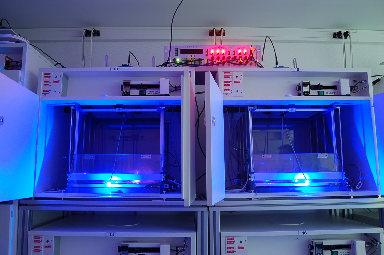
Image details
Original file; no crop, padding, color adjustment or depicted-content alteration.
Even the light itself needs a control. A 2019 study by Scott Owen, Max Liu and Anatol Kreitzer found that commonly used illumination conditions could warm brain tissue and alter firing in cells without an introduced opsin. In mice, illumination of the striatum could also bias turning behavior. An apparent response to an optogenetic intervention could therefore contain an effect of temperature.[11]
The researchers measured modest temperature rises, roughly 0.2 to 2 degrees Celsius under the conditions examined, and connected suppression of firing to a temperature-sensitive electrical conductance. Their finding was a practical warning about experimental design. A convincing result requires comparisons that distinguish the intended protein-mediated response from effects produced by the illumination apparatus itself. The power, duration and pattern of light delivery are part of the evidence, alongside the identity of the cells.[11]
From an experimental tool to a treatment
The possibility of interrupting harmful activity gave the technique medical appeal early on. In 2013, Esther Krook-Magnuson and colleagues tested a system that detected spontaneous seizures in a mouse model of temporal lobe epilepsy and responded with optogenetic stimulation. They could stop seizures either by inhibiting excitatory principal cells or by activating a small population of inhibitory neurons.[12]
The timing was central to the idea. A treatment delivered only when seizure activity began might avoid continuously interfering with the rest of the brain. The study joined detection and intervention into a feedback loop, showing that selected cell populations could alter an ongoing pathological event. But the result belonged to a mouse model, with the genetic preparation and implanted hardware that made the experiment possible. It supplied a direction for therapeutic research, not evidence that the same procedure was ready for patients.[12]
The eye has provided a concrete human example. In 2021, José-Alain Sahel, Botond Roska and collaborators reported partial recovery of visual function in a person with retinitis pigmentosa. They delivered genetic instructions for a light-sensitive protein into one eye, then used engineered goggles that converted changes in the visual scene into pulses of light directed at the retina.[13]
With the treated eye and goggles together, the participant could perceive, locate, count and touch different objects. Without the goggles after treatment, he could not detect the objects in those tests. The distinction is essential: the reported result depended on a combination of biological treatment and an external device. It was partial function in one person, not restoration of normal sight. The accompanying recordings of brain activity added evidence that the visual task was engaging the brain’s visual system.[13]
Clinical research has since explored other designs. A phase 2 study published in 2025 tested MCO-010 in six adults with Stargardt disease, a different inherited retinal condition. The treatment targeted surviving bipolar cells and was intended to make use of ambient light. The trial was open-label, followed participants for 48 weeks and was funded by Nanoscope Therapeutics.[14]
Average visual-acuity scores improved, but the small study had no randomized control group. All six participants had eye-related adverse events during follow-up; one had severe visual decline. No adverse event led to study discontinuation. Several authors disclosed company financial interests; sponsor personnel also participated in analysis and interpretation. The paper supported further investigation, including stronger controlled testing. It should be read as an early clinical result rather than as a promise that one injection will restore useful vision for everyone.[14]
For Deisseroth, the broader value still begins with discovery. Speaking to Stanford after the award, he described identifying the cells involved in a symptom as a step that can guide many kinds of intervention. A future treatment need not use light simply because light helped reveal the target. The Nobel recognizes a way of asking more precise questions of living tissue. Turning each answer into a dependable therapy remains another experimental task.[3]
Sources & further reading
Original reporting and research behind this article.
- Nobel Assembly: the 2026 Nobel Prize in Physiology or MedicineSource dated 2026-10-05; checked October 5, 2026
- Edward Boyden: a history of optogeneticsSource dated 2011-05-03; checked October 5, 2026
- Stanford: Karl Deisseroth wins the 2026 Nobel PrizeSource dated 2026-10-05; checked October 5, 2026
- Hegemann and Nagel: from channelrhodopsins to optogeneticsSource dated 2013-01-22; checked October 5, 2026
- Channelrhodopsin-1: a light-gated proton channel in green algaeSource dated 2002-06-28; checked October 5, 2026
- Channelrhodopsin-2, a directly light-gated cation-selective membrane channelSource dated 2003-11-13; checked October 5, 2026
- Millisecond-timescale, genetically targeted optical control of neural activitySource dated 2005-08-14; checked October 5, 2026
- Multimodal fast optical interrogation of neural circuitrySource dated 2007-04-05; checked October 5, 2026
- Millisecond-timescale optical control of neural dynamics in the nonhuman primate brainSource dated 2009-04-30; checked October 5, 2026
- Optogenetic stimulation of a hippocampal engram activates fear memory recallSource dated 2012-03-22; checked October 5, 2026
- Thermal constraints on in vivo optogenetic manipulationsSource dated 2019-06-17; checked October 5, 2026
- On-demand optogenetic control of spontaneous seizures in temporal lobe epilepsySource dated 2013-01-22; checked October 5, 2026
- Partial recovery of visual function in a blind patient after optogenetic therapySource dated 2021-05-24; checked October 5, 2026
- STARLIGHT: phase 2 optogenetic therapy trial in Stargardt diseaseSource dated 2025-08-14; checked October 5, 2026
- Neural substrates of awakening probed with optogenetic control of hypocretin neuronsSource dated 2007-10-17; checked October 5, 2026
THE DAYBREAK / THE MORNING LETTER
A little more understanding,
every morning.
The day’s selected stories, the context behind them, and a deeper read. A considered start to your day, delivered to your inbox.
See what’s inside